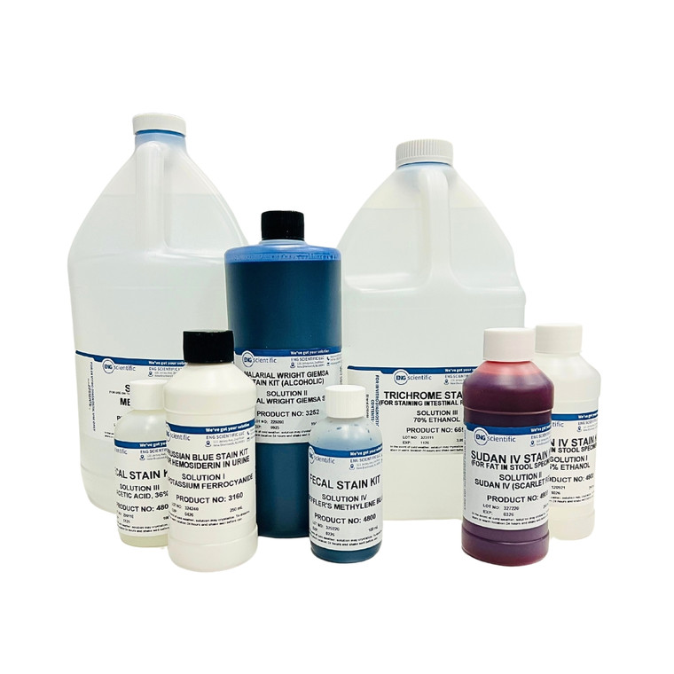
- Solution I - Aniline Blue
- Solution II - 0.5% Acid Fuchsin
Demonstrates collagenous fibrils, cartilage, bone, amyloid, and nuclei.
CONTENTS: Aniline Blue, Orange G, Phosphotungstic Acid, Acid Fuchsin
These solutions are made from certified dyes.
FOR IN VITRO DIAGNOSTIC USE ONLY.
RECOMMENDED PROCEDURE: Sections should be Zenker-fixed and procedure for removal of pigments and precipitates should be followed prior to staining. Stain with Sol. II for 1 - 5 minutes or longer. (Omit Sol. II to highlight collagenous fibrils). Transfer directly to Sol. I without washing in water. Stain for 20 - 60 minutes or longer. Dehydrate, clear and mount. For celloidin sections, shorten staining time, decolorize, dehydrate in 95% ethanol and clear by the blotting paper xylol method or in terpineol and mount in balsam.
RESULTS: Ground substances of cartilage and bone, mucus, amyloid - varying shades of blue; nuclei, fibroglia, myoglia and neuroglia fibrils, axis cylinders and fibrin -red; erythrocytes and myelin - yellow; elastic fibrils - pale pink, pale yellow or unstained.
Safety Data Sheet - Mallory's Aniline Blue Collagen Stain Kit (Solution I)
- UPC:
- 12352142
- Availability:
- 3-5 Days
- Type:
- Mallory's Aniline Blue Collagen Stain
- Includes:
- Aniline Blue, Orange G, Phosphotungstic Acid, Acid Fuchsin
- Format:
- Pack
- Hazmat:
- No
- WeightUOM:
- LB


